Soft X-ray microscopy (SXM)
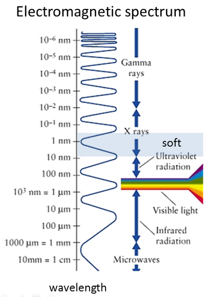
 .. uses electromagnetic radiation of 240 to 1.800 eV with wave lengths in the order of 7 nm to 0.7 nm, defining the physical limit of resolution.
... was developed in the 70ties by the group of Prof. Schmahl in Göttingen and is a powerful tool to image structures of solids by using high brilliant synchrotron radiation. In combination with spectroscopic methods it provides element specific information on the density distribution of local chemical, electronic and magnetic structure of solids.
... offers new routes to address a variety of static and dynamic issues in modern solid state science including material science, physics, chemistry and biology due to the unique simultaneous combination of important properties:

- High penetration depths of 100 nm to some µm
- High resolution down to 9 nm (best case)
- High temporal resolution down to 10 psec
- Significant chemical sensitivity Large magnetic cross sections
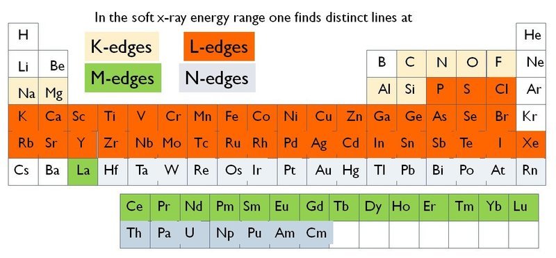
In the soft x-ray range nearly any element exhibit a characteric absorption edge.
