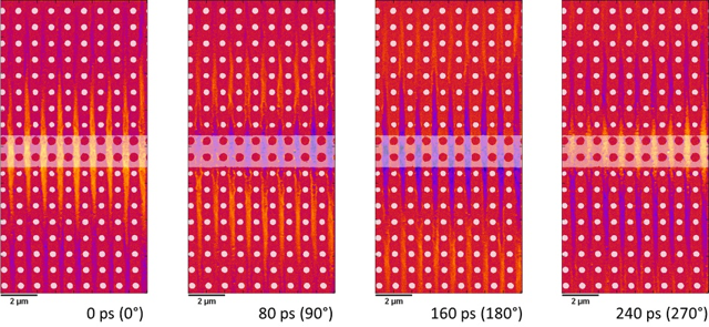
Artificial crystals for magnons or spin waves are imaged by high resolution time resolved x-ray microscopy. Spin waves are systematically influenced in these magnonic crystals and a spin wave band structure is formed for different spin waves, similar to photonic crystals for photons. The complex interplay of different spin wave types can only be directly imaged by scanning x-ray transmission microscopy (at the MAXYMUS end station at the BESSY II storage ring in Berlin) with high temporal (<40 ps) and spatial (<20 nm) resolution. These experiments result in a combined amplitude and phase information of all spin waves in the system and yield a unique access to the fundamental spin wave properties and allow a substantial understanding of magnonic crystals. Building on this understand complex magnonic structures can be constructed.

Dynamic x-ray micrographs with magnetic contrast (XMCD) at different time steps of a video that shows the propagation of spin waves in an antidote lattice. Here, spin waves can only propagate between holes. The applied bias field allows for a propagation of more than 10 µm away from the excitation source.
