Special SXM Techniques
One of our core capabilities is the development of cutting edge SXM techniques that are not available other end stations. With a dedicated team of engineers and scientists we drive SXM imaging beyond widespread limitations in terms of sample environment, signal detection and imaging capabilities. These efforts include conventional hardware and software development as well as exploring and leveraging new fundamental physics for improving x-ray imaging.
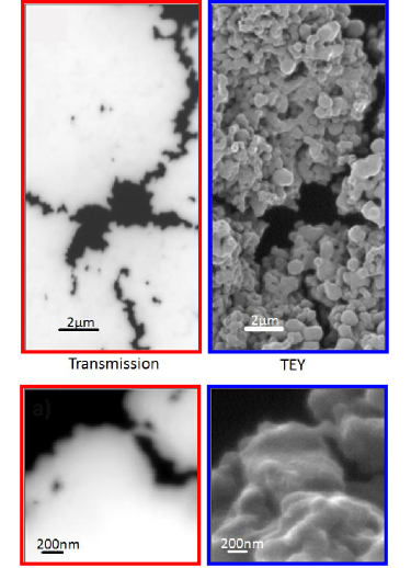
Comparison of transmission and TEY mode
 Due to the UHV condition the absorption of the x-rays can be also monitored via detection of the total electron yield (TEY). This method, which provides a quasi-3D image similar to SEM pictures, is inherently strongly surface sensitive probing a depth of 1 to 3 nm as demonstrated by a study of agglomerated FexOy/PtFe hybrid particles. By combining TEY and transmission mode differences of the chemical characteristics of the surface and the bulk can be separated.
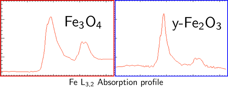
In the transmission mode the profile of the absorption spectra are more likely Fe3O4 like. The corresponding TEY absorption spectra prove , that the surface chemical
characteristic is y-Fe2O3 like

Ptychography
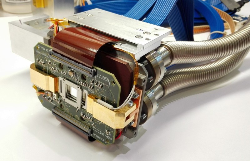
 Ptychography is an image enhancement technique that can be implemented for scanning microscopes. In simple terms, it relies on replacing the point detector with an area detector behind the sample. In our case, this means replacing the APD detector with a fast x-ray CCD camera. Thus, in addition to the transmitted intensity, the x-ray scattering pattern after transmission through the sample is collected. This additional information allows increasing the resolution and signal-to-noise ratio beyond the capabilities of the x-ray optics that are used and allows us to reach single digit nanometer resolutions. To leverage the information contained in the recorded scattering patterns, a recursive reconstruction algorithm is needed that is executed on a dedicated GPU computation node. This high-performance setup allows online reconstruction of the measurement data. Ptychographic image enhancement in combination with the high photon flux at the MAXYMUS beamline allows us to push the resolution and contrast envelope to allow ultimate x-ray imaging.
